Important Neurological Assessments

Getting the neurological picture
- Medical and family history
- Developmental history
- Brain Trauma
- Growth parameters assessment
- Cognitive assessment
Mental health and behavior evaluation - Motor function and muscle tone assessment
- Sensory function
- EEG, MRI, CT Scan
- Assessments of cerebrospinal fluid (CSF)
- Genetic testing
A good neurological assessment includes:
- Pupillary reaction
- Motor skills
- Consciousness
- Cranial nerves and swallowing/Gag ability
- Coordination
- Reflexes
- Perception
- Language and cognition
- Vital signs: Abnormal breathing, for example may occurs in brain injury.

The NCLEX loves these two postures which occur in severe neurological damage
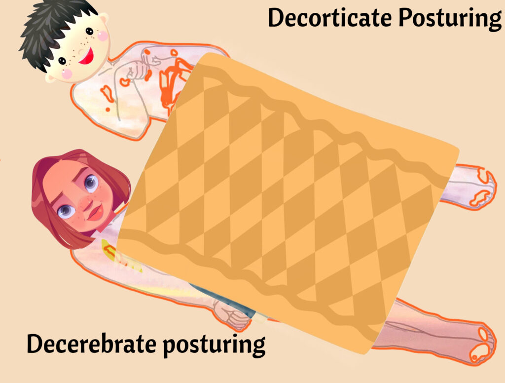
Decorticate: notice the FLEXION of the arms toward to cor (center of the body). deCORticate
Decerebrate: notice the EXTENSION of the arms. dEcErEbratE has lots of “E’s” like ExtEnsion.
Newborns have several reflexes that are critical for their survival and development. These reflexes are automatic and involuntary responses to certain stimuli. They include:
- Sucking reflex – This occurs when a baby’s lips are touched, causing them to instinctively suck, even if nothing is in their mouth. It usually disappears at around 3-4 months.
- Rooting reflex – When a baby’s cheek is touched, they will turn their head towards the touch and make sucking motions with their mouth. This reflex disappears at around 4 months.
- Grasping reflex – If an object is placed in a baby’s palm or foot, they will instinctively grasp it. This reflex usually disappears at around 3-4 months.
- Moro reflex – If a baby is startled or feels like they are falling, they will throw their arms and legs out and then bring them back in. This reflex disappears at around 3-4 months.
- Babinski reflex – When the bottom of a baby’s foot is stroked, their toes will spread out. This reflex disappears at around 12 months.
Spinal Cord Damage
The upper motor neuron descends in the spinal cord to the spinal nerve root. The upper motor neuron synapses with the lower motor neuron in the ventral horn of the spinal cord. The lower motor neuron then innervates the skeletal muscle.
Lower Motor Neuron
Lower motor neuron lesions present with muscle atrophy, fasciculations (muscle twitching), decreased reflexes, decreased tone, and flaccid paralysis. Think Lower = Low tone (flaccid paralysis), low reflexes, and atrophy (low muscle tone)
Upper Motor Neuron
Upper motor neuron lesions present with weakness, spasticity (spastic paralysis), clonus, and hyperreflexia. Upper = high tone, hyper (high reflexes) and spastic (high tone paralysis),
Upper motor neuron damage: cerebral palsy, traumatic head injuries, and spinal cord injuries.
Lower motor neuron damage: muscular dystrophy, spinal muscular atrophy, and peripheral nerve injuries.
Assessing for Meningitis
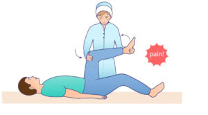
Kernig Sign = Knee Extension is painful
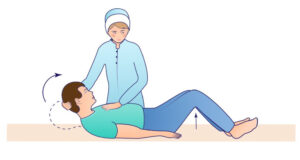
BrudziNsKi Sign = Neck Flexion leads to Knee Flexion
Meningitis is a serious and potentially life-threatening infection of the protective membranes that cover the brain and spinal cord. The signs and symptoms of meningitis include fever, headache, nausea or vomiting, stiff neck, sensitivity to light, confusion, and seizures.
Other signs that may indicate meningitis include a rash of tiny, red or purple spots that appear anywhere on the body, irritability, drowsiness, and difficulty waking up. In infants, meningitis may present with a bulge in the soft spot on the top of the head, a high-pitched cry or moaning, and poor feeding.
The nurse performs a pediatric neurological assessment on a child with a mild head injury. Which of the following would the nurse not include in this assessment?
The nurse is caring for a 12-month-old child. Which reflex may still be present in this child?
All the other reflexes disappear around 3 to 6 months. Babinski reflex may be present up 12 months or a little longer.
During a pediatric neurological assessment, what is the nurse looking for with the assessment of Kernig's sign in a child with suspected meningitis?
Kernig's sign is used to diagnose meningitis
The nurse anticipates which of the following in a child with upper motor neuron damage?
This is important to know: Upper motor neuron damage= weakness, spasticity (spastic paralysis), clonus, and hyperreflexia. Lower motor neuron lesions present with muscle atrophy, fasciculations (muscle twitching), decreased reflexes, decreased tone, and flaccid paralysis. Positive Romberg's test is an inability to maintain an erect posture over 60 seconds with eyes closed (important in multiple sclerosis).
The nurse expects which of the following findings in a child with a brain injury?
During a pediatric neurological assessment, what is the appropriate method for testing for nystagmus?
Nystagmus is a condition where the eyes move rapidly and uncontrollably. They can move: side to side (horizontal nystagmus), up and down (vertical nystagmus), in a circle (rotary nystagmus).
Which of the following is most likely to be a sign of increased intracranial pressure during a pediatric neurological assessment?
This occurs when a baby’s lips are touched, causing them to instinctively suck, even if nothing is in their mouth. It usually disappears at around 3-4 months.
When the bottom of a baby’s foot is stroked, their toes will spread out. This reflex disappears at around 12 months.
The nurse is caring for a client with upper motor neuron damage. The nurse expects which of the following symptoms?
A client with upper motor neuron damage would likely demonstrate which of the following on physical examination?
A client with lower motor neuron damage would likely demonstrate which of the following on physical examination?
Which region of the brain is primarily responsible for upper motor neuron function?
I did not tell you about this. Did you choose the right answer. We are talking about motor neurons so motor cortex makes sense.
A 6-year-old male child presents to the clinic with complaints of muscle weakness, difficulty walking and speaking. On examination, the child shows a waddling gait, difficulty standing on heels and has a positive Gower sign. Which condition is most likely causing these symptoms?
One of my students had this quesion on her NCLEX, so I just wanted to surprise you. Duchenne Muscular Dystrophy (DMD) is a genetic disorder that affects the muscles. Children with DMD have decreased muscle strength and progressive muscle weakness, which leads to difficulty walking and eventually requires a wheelchair.