Heart Failure
4 Topics
EKG Rhythms
2 Topics
Anatomy and Physiology
Here is some anatomy and physiology
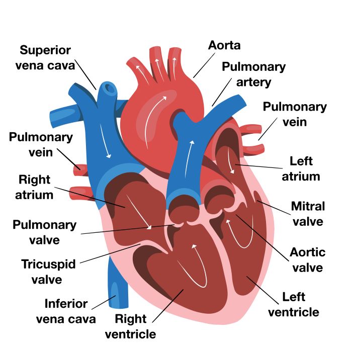
Please know...
- Vessels from the heart to the body are arteries. Right? And vessels that bring blood back to the heart are veins. Right?
- The superior and the inferior vena cava bring blood from the body to the right side of the heart.
- The pulmonary artery, brings takes blood from the right side of the heart to the lungs, is the only artery with deoxygenated blood or venous blood. Think about it. It is an artery because it takes blood from the heart to the body (or lungs in this case).
- The pulmonary veins are the only veins that carry 100% oxygen or oxygenated blood. There are veins because they carry blood from the lungs to the heart (body to heart vessels so veins!).
Hotspot: Click on the expand buttom on the right upper corner to expand and see the heart on the full screen
The three layers of the heart wall
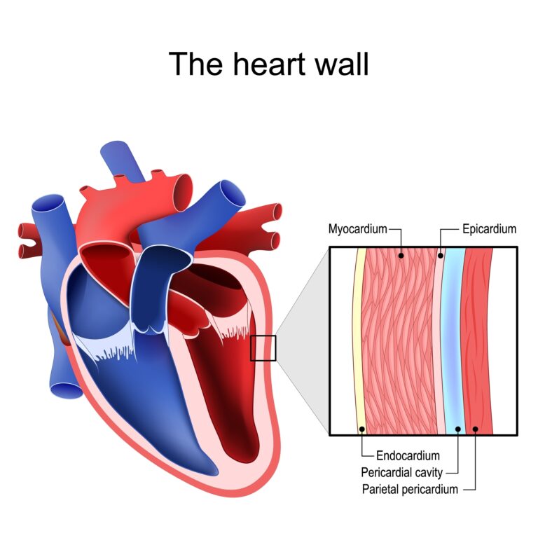
- Epicardium: outermost layer
- Myocardiam: middle layer
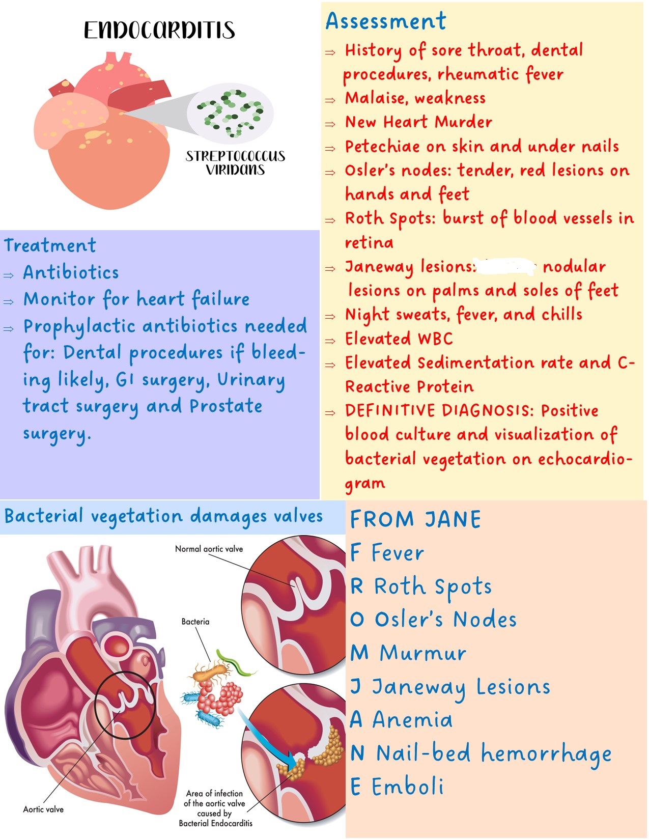
- endocardium: inner layers that lines inner chambers and heart vales. Remember that endocardium lines heart valves. This is important because endocarditis may lead to valve damage.
Pericardial Sac
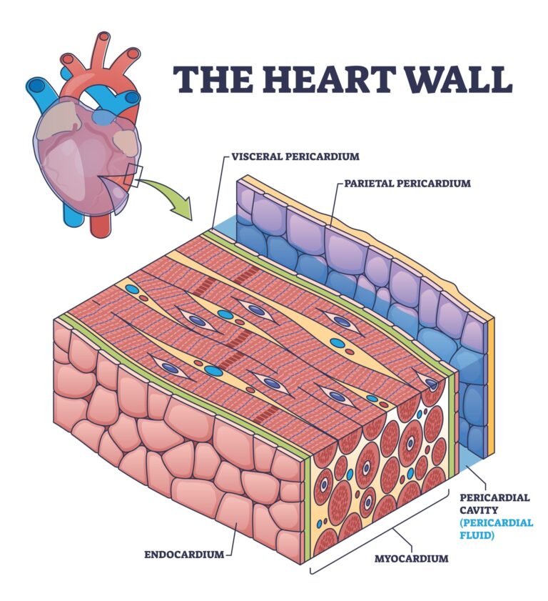
- Surrounds the heart and protects it from trauma and infection
- It has two layers: the parietal pericardium and the visceral pericardium
- Parietal pericardium is the tough fibrous outer membrane
- The visceral pericardium is the thin, inner layer (To remember let’s think this way: visceral=inner organ. Visceral attaches to the organ, in this case the heart).
- The pericardial space is between the parietal and visceral layer. It holds about 5 to 20 ml of pericardial fluid. This fluid lubricates pericardial surfaces and cushions the heart.
The pericardium can either become inflamed (infection) or fill with fluid (effusion)
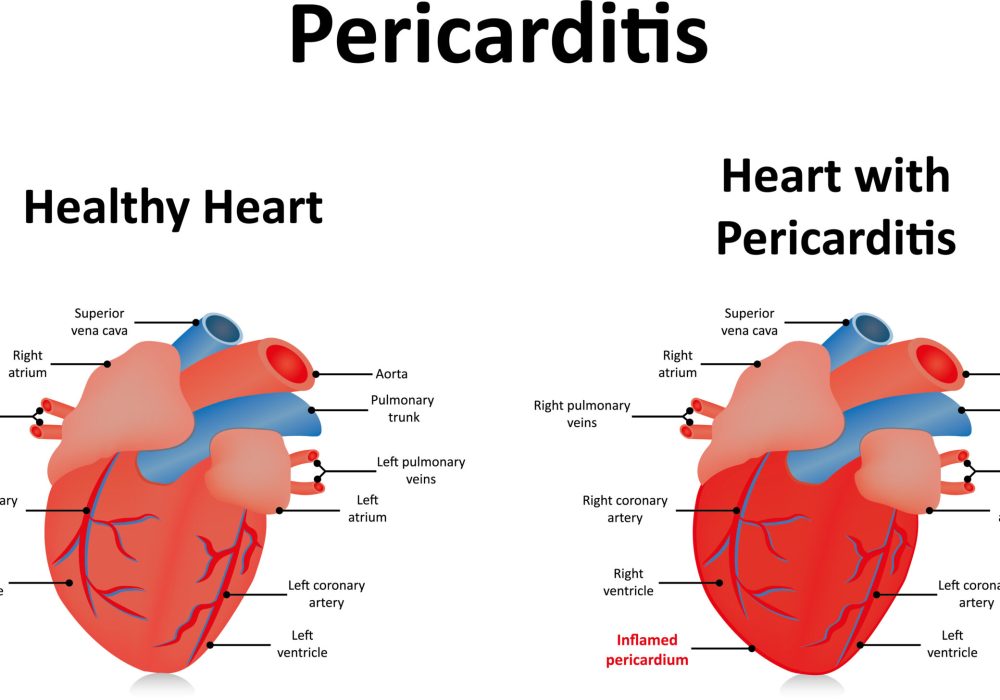
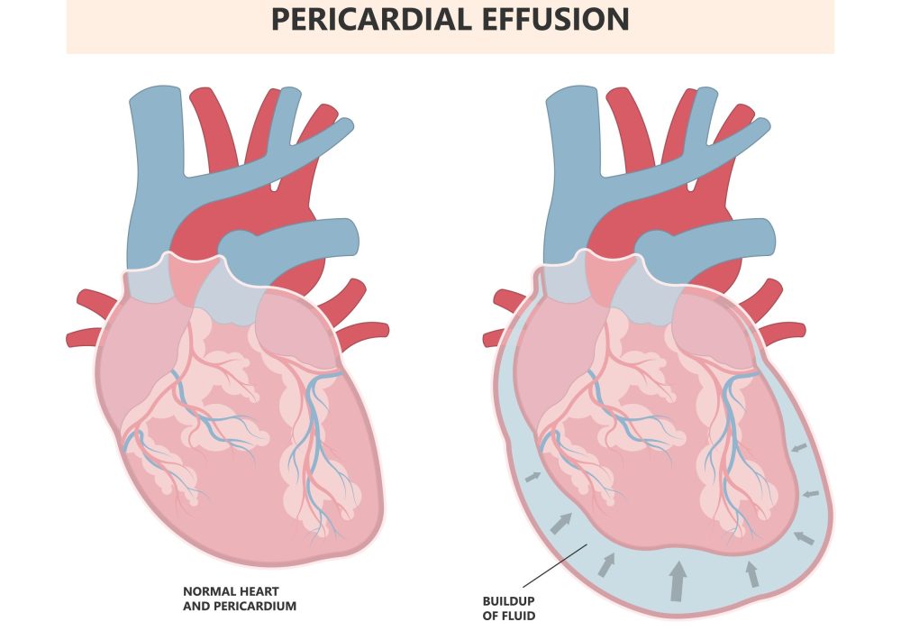
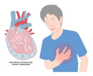
Which can lead to cardiac tamponade- pressure on the heart preventing it from contracting. Poor heart contraction = abnormal vital signs and poor organ perfusion
Here are the chambers of the heart
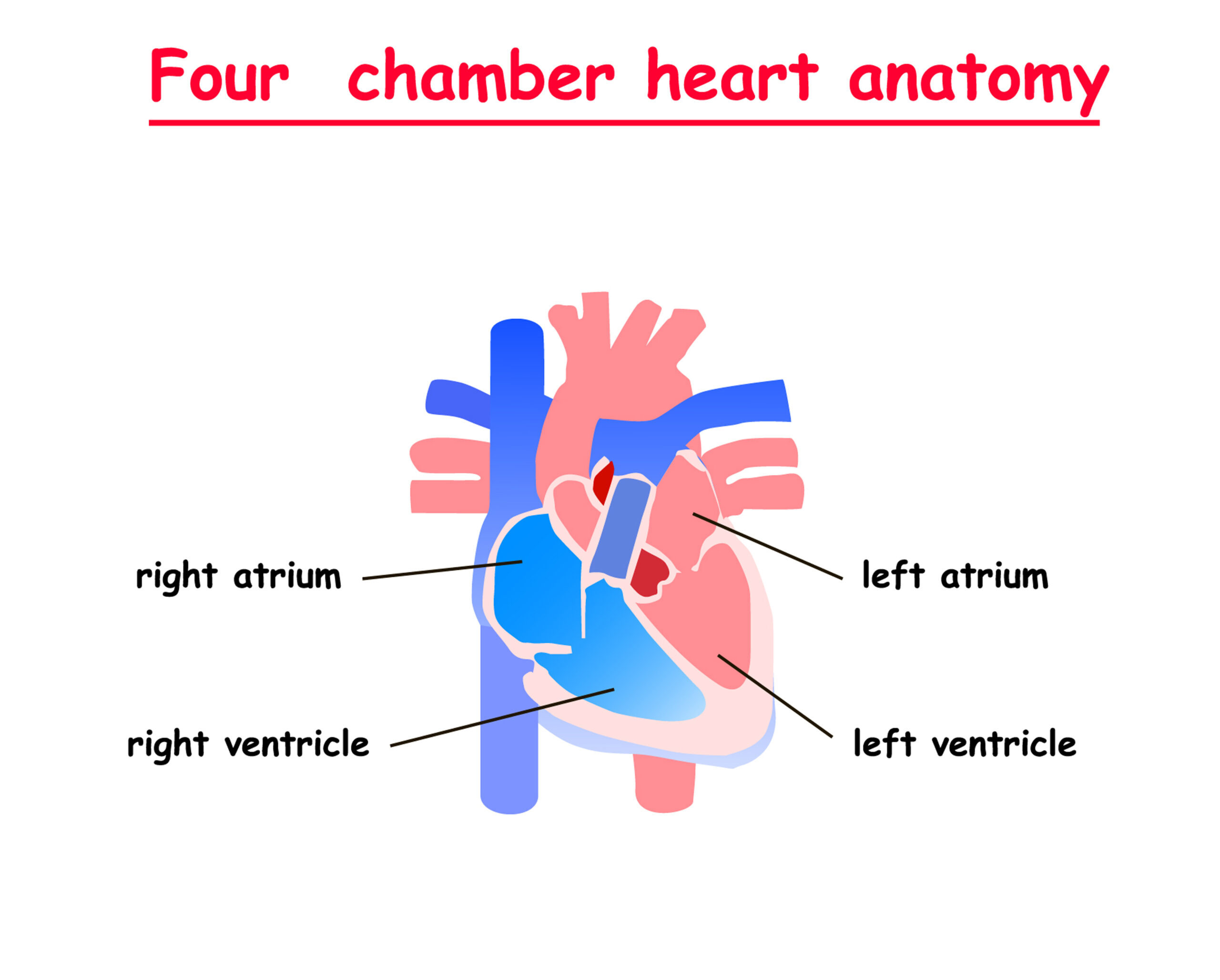
Simple right! The right side carries the venous blood coming from the body (designated as blue) and the left side carries the arterial blood that comes from the lungs to the body (designated as red).
A little more detail...
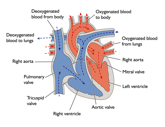
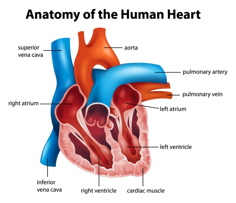
- Veins come from the body and go to the heart and arteries go away from the heart towards the body.
- Veins carry deoxygenated blood
- Arteries carry oxygenated blood
- Two big exceptions: The pulmonary artery and the pulmonary veins.
- The pulmonary veins carry oxygenated blood because they are carrying the blood that comes from the lungs.
- The pulmonary artery carries deoxygenated blood because it carries the blood that comes from the body to the lungs where blood will become oxygenated.
Always remember the paradox of the pulmonary artery and the pulmonary veins.
The four cardiac valves
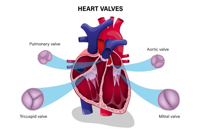
- Tricuspid is between the right atrium and the right ventricl
- Think TRISCUPID=TRINITY. Tricuspid is on the right because the church is always right. Silly way to remember, I know.
- The valves between the atria and the ventricles are the tricuspid and the mitrai valve
- The tricuspids has three cusps and mitral has only two (bicuspid)
- Location of the aortic and the pulmonary valve should be self-explanatory at this point. They are usually called semilunar valves.
Everybody always wonders when do valves open and when do they close...
- Let’s think through it. When the heart contracts blood needs to be ejected to go to the organs. For this to happen aortic and pulmonary valve must be open.
- So systole (contraction) Aortic and Pulmonary valve are open and diastole (relaxation) when the heart is filling, of course they are close to keep the blood in the heart.
- Tricuspid and mitral do the opposite. Open during diastole to allow heart to fill and close during systole.
Systole- Pulmonary and Aortic valve Open to eject the blood
Diastole- Tricuspid and Mitral valve Open to fill the heart
Let's see it in action!
The electrical conduction of the heart
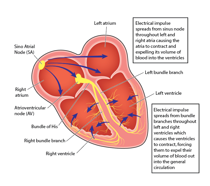
SA node is the main pacemaker of the heart. It generates impulses at 60 to 100 beats/minute
If SA node fails, AV node can take over, but only at 40 to 60 beats/minute. Ask yourself, would that be enough to maintain cardiac output and tissue perfusion. Usually not.
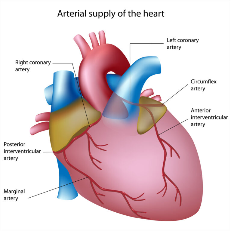
The Coronary Arteries
